Methodology
Table of contents
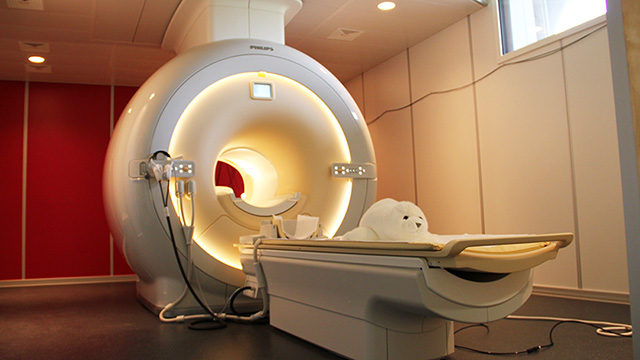
Magnetic resonance imaging (MRI)

Magnetic resonance imaging (MRI) is an extremely versatile imaging method. It allows us to take pictures of the brain. It utilises the different magnetic properties of particles in different tissues of the brain. For instance, the different tissues in the brain can be visualised to allow for a detailed examination of the brain anatomy. Furthermore, special MRI variants can be utilised to examine blood vessels, metabolites and fibre bundles.
Watch our movie about MR Imaging
Where does our brain work?
Functional MRI (fMRI) can be used to investigate brain function. The participants solve certain tasks while the MRI device records brain images in rapid succession. Using complex arithmetic operations, we can calculate from the recorded images which brain regions are active during a specific task (e.g. reading) and how they work together in networks (connectivity).
Why research with fMRI?
Functional magnetic resonance imaging (fMRI) provides insights into brain processing that cannot be captured by behavioural studies alone. By comparing patients and healthy test subjects, we can identify impaired brain networks. The comparison between children and adults allows us to study the development and maturation of the brain. A major goal of our research is to better understand the brain and identify factors that can improve diagnosis and prediction as well as contribute to the development of new, targeted interventions.
Is MRI dangerous?
MRI is a commonly employed diagnostic tool in clinical settings, as well as in research involving subjects of all ages, from infants to the elderly. It is not a dangerous procedure. Because the MRI machine emits a loud noise, all participants are provided with hearing protection. In contrast to computer tomography, MRI does not require X-rays to produce images.
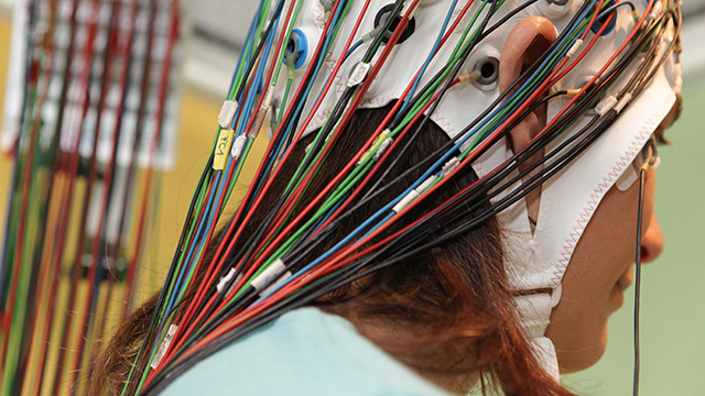
Electroencephalography (EEG)

Electroencephalography (EEG) is a neurophysiological method that provides a detailed insight into the temporal processing of information in the brain. Sensors (electrodes) on the surface of the head are used to measure the smallest electrical voltages generated by the activity of the nerve cells in the brain. The method does not permit the measurement of the activity of individual cells; rather, it provides an indication of the combined activity of a large number of cells in the brain. The data can be presented in the form of curves (over time) or "maps".
The spontaneous EEG
The spontaneous EEG is composed of oscillations of different frequencies.
It has been demonstrated that certain frequency bands are more prevalent in specific states of consciousness. Alpha waves (8-12 Hz) are typically observed in a relaxed state with eyes closed. However, the frequency band composition also undergoes changes with development and is altered in certain diseases.
Event-related potentials (ERP)
Event-related potentials (ERP) are observed with a constant temporal reference to an event (e.g. image, sound, etc.). They are typically quite weak and are superimposed by the larger spontaneous electroencephalogram (EEG). However, ERPs become visible through a large number of repetitions of the same stimulus and subsequent averaging. This process allows for the random (i.e. non-event-related) activity to be averaged out, leaving only the event-related activity, which becomes visible as an ERP.
ERPs exhibit characteristic latencies and potential distributions. Comparative analysis of ERPs between children and adults or between patients and healthy individuals can provide insights into the development of specific processes and the impairment of those processes in patients.
Is EEG a dangerous procedure?
EEG is a safe and non-invasive procedure. It is therefore widely used in both routine clinical diagnostics (e.g. for epilepsy) and research studies involving infants, children, adolescents and adults.
An example from reading research
An example from the field of reading research is the use of electroencephalography (EEG) to examine the highly specialized processing of writing in the brain. EEG data reveals that writing is processed differently in the brain than "meaningless" symbols, despite the visual similarity between words and symbol sequences. This distinction becomes apparent as early as 150-200 milliseconds after a word is presented. The ERP "N1" displays a potential distribution with a strong negativity over the left back of the head for words. This negativity is much less pronounced for series of symbols. Additionally, a difference has been identified in this ERP between children with reading difficulties and children without reading difficulties. This suggests that writing is processed differently, presumably less efficiently, in children who have problems with reading. However, the underlying cause of this remains unclear.
Combined EEG-fMRI imaging
The combination of EEG and fMRI imaging represents a significant advance in the field of neuroimaging. The advent of new technologies has made it possible to perform simultaneous EEG and fMRI imaging, which offers a more comprehensive understanding of the brain's functional activity.
In combined EEG-fMRI imaging, the subject is positioned inside the MRI machine with an EEG cap on the scalp. The brain waves (EEG) and the brain images are recorded simultaneously. In this manner, the advantages of both methods are combined in a single session. The fMRI enables the identification of which brain regions are active during a particularc task, while the event-related potentials of the EEG provide information about the temporal course of information processing.